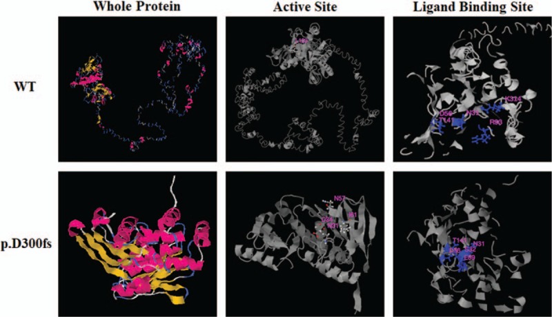
Figure 5.

Prediction of the tertiary structure and function for wild type PMS1 and the p.D300fs mutant proteins with I-TASSER tool (https://zhanglab.ccmb.med.umich.edu/I-TASSER/). The tertiary structure (column ‘whole protein’), the prediction of enzyme active site (column ‘active site’) and the prediction of ligand binding site (column ‘ligand binding site’) of the both WT and mutant protein are shown as indicated.
