
. 2019 Sep 17;69(1):80–91. doi: 10.1538/expanim.19-0079
©2020 Japanese Association for Laboratory Animal
Science
This is an open-access article distributed under the terms of the Creative Commons Attribution Non-Commercial No Derivatives (by-nc-nd) License. (CC-BY-NC-ND 4.0: https://creativecommons.org/licenses/by-nc-nd/4.0/)
Fig. 4.
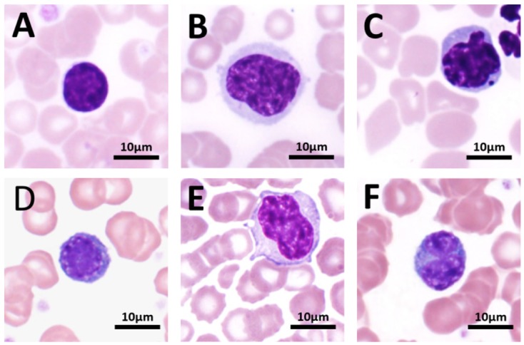
Various lymphocytes under a light microscope (100×).