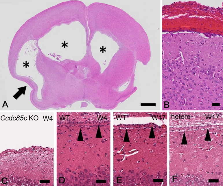
Fig. 3.
Histopathological analysis of hydrocephalus in Ccdc85c KO rats. HE stained sections of cerebrum in Ccdc85c KO rats at four weeks of age (4W) (A–C) and unaffected rats at 4W [D: wild type (WT)] or at 17W (E: WT, F: hetero). Lateral ventricles are dilated in Ccdc85c KO rats (A, asterisks). Atrophied cerebral parenchyma is also observed (A, arrow). Hemorrhage is mainly located at meninges (B). Lateral ventricles are lined by ependymal cells in WT rat and hetero rat (D–F, arrowheads). There are no ependymal cells at the lateral ventricles in Ccdc85c KO rats (C). Scale bar of A, 1 mm, scale bar of B, 50 µm, scale bar of C–F, 25 µm.

