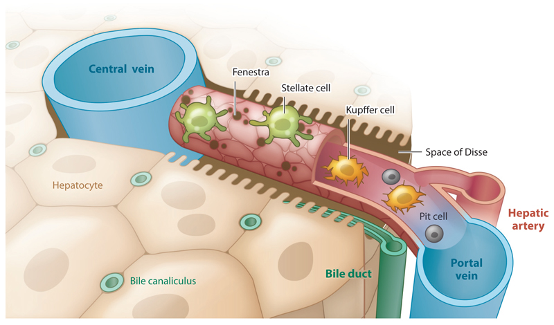
Figure 1.
Schematic of the hepatic sinusoid. The sinusoid is a fenestrated capillary, 275 μm in length, composed of sinusoidal endothelial cells (red). The liver-resident pericytes, called stellate cells (green), wrap the capillary. Resident macrophages, called Kupffer cells (yellow), and natural killer cells, called pit cells (gray), roam the inside of the vessel.

