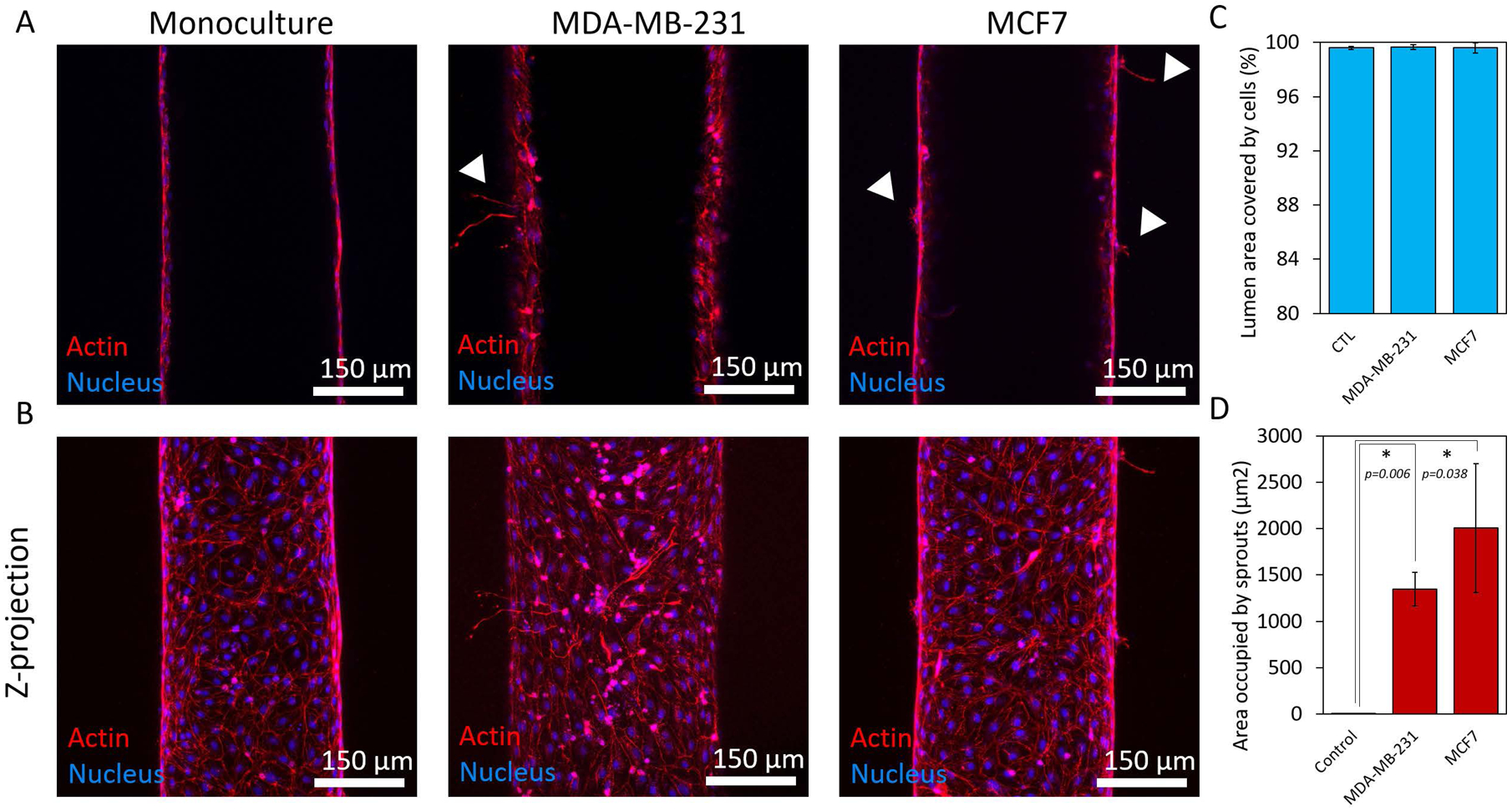
Figure 3.

Endothelial cell coverage and lymphangiogenic sprouting. A) Confocal images showing the middle plane of lymphatic vessels in monoculture and co-culture with MCD7 and MDA-MB-231 cells. B) Z-projected images of vessels in monoculture and co-culture with MCF7 and MDA-MB-231 cells. C) Quantification of cell coverage for each culture condition showing an average percentage of cell coverage >99% for all conditions. D) Crosstalk with the breast cancer cells induced lymphangiogenic sprouting in the vessels. There were no observable sprouts in the monoculture control. Three individual vessels (n = 3) were measured for each culture condition to determine the average permeability value (mean ± s.d.).
