Figure 2.
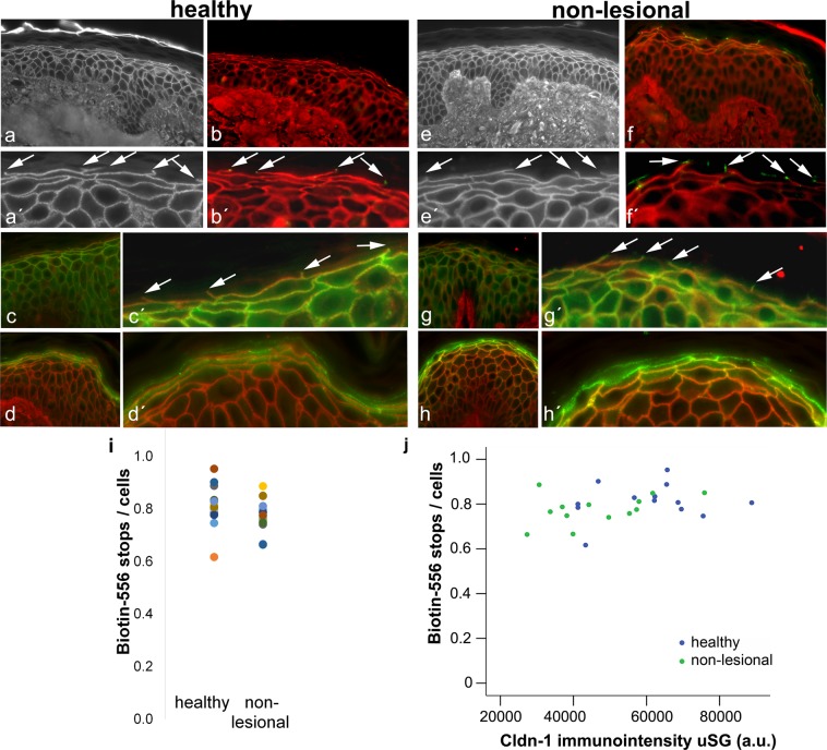
TJ barrier in healthy and AD non-lesional skin and its correlation to Cldn-1 levels. (a,a′,e,e′) Examples for Biotin-556-TJ barrier assays in healthy (a,a′) and non-lesional AD (e,e′) skin. (b,b′,f,f′) Double-staining of Biotin-556 (red) and occludin (green), (c,c′,g,g′) of Biotin-556 (red) and Cldn-1 (green), and (d,d′,h,h′) of Biotin-556 (red) and Cldn-4 (green) in healthy (b,b′,c,c′,d,d′) and non-lesional (f,f′,g,g′,h,h′) skin. (a′,b′,c′,d′,e′,f′,g′,h′) are magnifications of (a–h). Arrows denote “tracer-stops”. (i) Number of tracer stops per number of cells in the underlying layer in healthy and AD non-lesional skin. Each dot represents a proband/patient (n = 13 per group). (j) Correlation of Biotin-556 tracer stops/cells with Cldn-1 immunointensity in SG in healthy and non-lesional AD skin. Blue dots: healthy, green dots: non-lesional.

