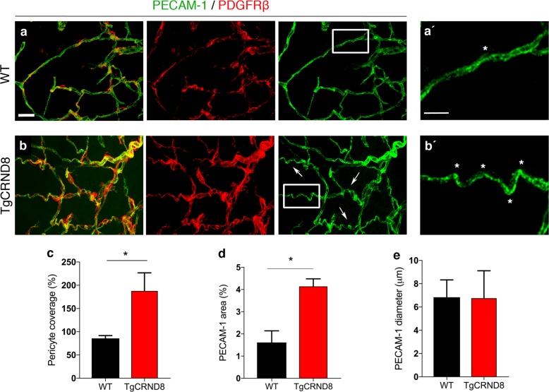
Fig. 1. Vascular morphology in the cortex of postnatal TgCRND8 mice.
a, b Blood vessels were stained by PECAM-1 and pericytes by PDGFRβ. Increased pericyte coverage and microvessel tortuosity (arrows and framed areas) is seen in P7 TgCRND8 cortex; scale bar 20 μm. (a′, b′) Enlarged white-framed areas in a and b showing samples of normal microvessels in WT (a′) and tortuous microvessels (*) in TgCRND8 (b′) cortex; scale bar 10 μm. c There is increased PDGFRβ+ pericyte coverage in TgCRND8 cortex (*p < 0.05). d There is an increase in PECAM-1+ area in TgCRND8 cortex (*p < 0.05). e No difference in vessel diameter was detected (p > 0.05); mean ± SD (n = 2 (WT), n = 4 (TgCRND8), Student’s t-test.

