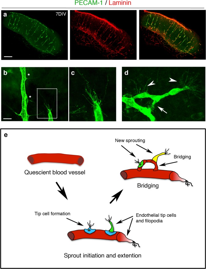
Fig. 2. Characterisation of sprouting angiogenesis in 7-days in vitro organotypic cortical slice cultures from wild-type mice.
a Confocal images of cortical slices stained for PECAM-1 (green) and laminin (red) to visualise blood vessels at 7 days in vitro, scale bar 500 μm. b–d Different stages of vascular sprouting can be visualised in cortical brain slices including; tip cell formation (*) endothelial tip extension (framed area in b which is expanded in c) new sprouting (arrow heads) and bridging (arrow) (scale bar 50 μm) (d). e Diagram summarising the different stages of sprouting angiogenesis that can be observed in cortical organotypic slice cultures.

