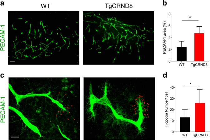
Fig. 4. TgCRND8 organotypic cortical slices show increased vascular density and excessive filopodia formation when compared to wild-type cultures.
a Representative confocal images showing blood vessel density (PECAM-1, green) in 7 days in vitro WT and TgCRND8 slices; scale bar 100 μm. b Quantification of PECAM-1+ area (as percentage of the total image) reveals a significantly higher blood vessel density in TgCRND8 cortical slices vs. WT slices. (mean ± SD (n = 5 (WT), n = 4 (TgCRND8), *P < 0.05 Student’s t-test). c Confocal images showing endothelial cells extending numerous finger-like filopodia at the forefront of vascular sprouts in 7 days in vitro WT and TgCRND8 cortical slices, visualised by PECAM-1 labelling. Red dots highlight vascular sprouting tips, scale bar 20 μm. d Quantification shows that the number of filopodia per cell is significantly higher in TgCRND8 when compared to WT slices. (mean ± SD, (n = 14 (WT), n = 13 (TgCRND8), *P < 0.05 Student’s t-test).

