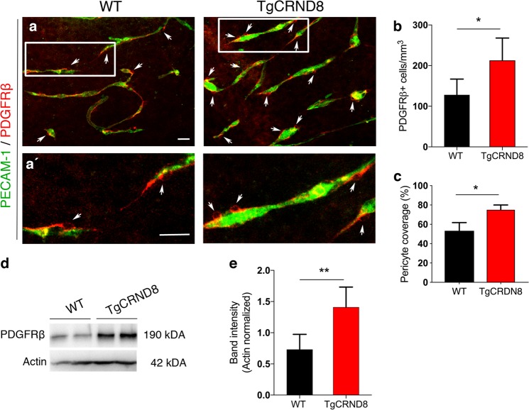
Fig. 5. Increased pericyte number and PDGFRβ expression in TgCRND8 organotypic cortical slices.
a Representative confocal images showing PDGFRβ+ pericytes (arrows) around blood vessels (PECAM-1, green) in 7 days in vitro WT and TgCRND8 slices; scale bar 50 μm. PDGFRβ+ pericytes are closely associated with cortical microvessels. a′ The framed areas in a are enlarged, scale bar 50 μm. b Quantification of PDGFRβ+ pericytes reveals an increased number in 7 days in vitro TgCRND8 slices when compared to WT (data are expressed as cell numbers per mm3. c Quantification of PDGFRβ+ staining area normalised to PECAM-1+ staining area shows a significant increase in pericyte coverage in TgCRND8 OBSCs; mean ± SD. (n = 4 (WT), n = 5 (TgCRND8), *P < 0.05 Student’s t-test). Representative western blots (d) and quantification of PDGFRβ band intensity (e) in 7 days in vitro WT and TgCRND8 cortical slices, shows increased PDGFRβ in TgCRND8 cultures when normalised to Actin control (n = 5 (WT), n = 4 (TgCRND8) **P < 0.01 Student’s t-test).

