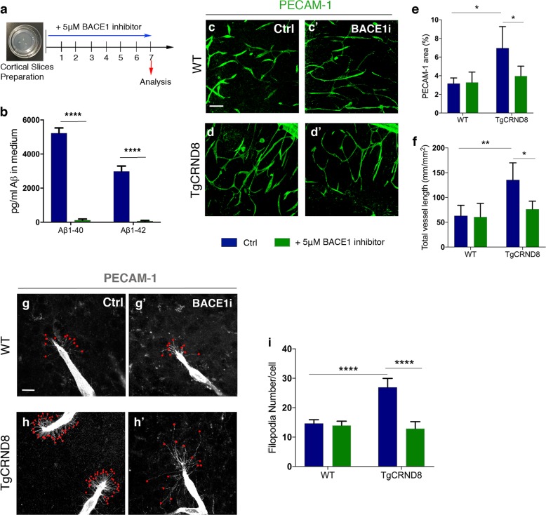
Fig. 6. BACE1 inhibition decreases vascular density and normalises aberrant vascular sprouting in TgCRND8 organotypic cortical slices.
a Diagram showing the experimental schedule for BACE1 inhibitor treatment of WT and TgCRND8 cortical slices. b Measurement of Aβ1–40 and Aβ1–42 in the culture medium of 7 day in vitro TgCRND8 slices treated with BACE1 inhibitor (mean ± SEM (n = 4), two-way ANOVA effect of treatment ****P < 0.0001). c Confocal images showing blood vessel density (PECAM-1, green) in 7 days in vitro control (c) and BACE1 inhibitor (c′) treated WT slices. d–d´ Confocal images showing blood vessel density (PECAM-1, green) in 7 days in vitro control (d) and BACE1 inhibitor (d′) treated TgCRND8 slices; scale bar 20 μm. BACE1 inhibition rescues the increase in PECAM1+ area (percentage image coverage) (mean ± SD (n = 4 (WT), n = 4 (TgCRND8), *P<0.05, two-way ANOVA) (e) and total vessel length (mm/mm2) (mean ± SD (n = 4 (WT), n = 4 (TgCRND8), **P<0.01 and *P < 0.05, two-way ANOVA) (f) seen in control treated TgCRND8 cultures. PECAM-1 staining of WT (g–g′) and TgCRND8 (h–h′) slice cultures with (g′, h′) or without (g, h) BACE1 inhibitor treatment. Individual filopodia are highlighted with red dots. i Quantification of the number of filopodia per cell demonstrates that BACE1 inhibitor rescues the increased number seen in untreated TgCRND8 cultures when compared to WT at 7 days in vitro (mean ± SD, (n = 4 (WT), n = 4 (TgCRND8), ****P<0.0001, two-way ANOVA).

