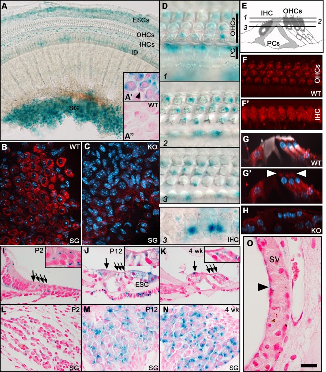
Fig. 1. MANF expression in the cochlea, revealed by X-gal histochemistry and immunostaining.
a The whole mount shows prominent X-gal staining in the SG, IDs, IHCs, and ESCs. The weaker staining in OHCs is not evident in this low magnification. a′ X-gal staining is present in SG neurons and absent in the satellite cells (arrowhead), revealed in a paraffin section. a″ X-gal staining is absent from SG neurons from a WT mouse. b MANF immunostaining (red) is prominent in SG neurons, revealed in a whole mount. DAPI marks nuclei (blue). c SG neurons from a KO mouse lack MANF staining. d Whole mount is viewed at different planes from the hair cell cuticular plate down to nuclear level. X-gal staining is prominent in IHCs and pillar cells (supporting cells). In OHCs, X-gal staining is concentrated to the plane below the cuticular plate (plane 2). Note, however, that X-gal staining is not a readout of intracellular protein distribution pattern. e Schematic cross-section through the organ of Corti depicts the imaging planes in d and the cell types. f Whole mount shows MANF immunostaining in OHCs, at the plane below the cuticular plate. f′ The same wholemount shows widespread MANF immunostaining in IHCs. g, g′ Maximum intensity projection of MANF-immunostained (red) whole mount viewed in transverse plane. Phalloidin (white) labels F-actin and DAPI (blue) nuclei. MANF is expressed below the cuticular plate (arrowheads) in OHCs. MANF is widely expressed in the IHC cytoplasm as well as in Deiters’ cells (supporting cells) beneath OHCs. Phalloidin labels the hair cell apices and pillar cells. h MANF immunostaining is absent from the organ of Corti of KO mice. i, j, k Transverse paraffin sections show X-gal staining in the organ of Corti at different ages. Large arrows mark IHCs, smaller arrows the three OHC rows. Insets show OHCs in higher magnification. l, m, n Transverse paraffin sections show the increase of X-gal-staining intensity along aging. o Manf is very weakly expressed in the luminally located marginal cells (arrowhead) of the stria vascularis. Abbreviations: KO knock out; WT wildtype; SG spiral ganglion; OHCs outer hair cells; IHCs inner hair cells; IDs interdental cells; ESCs external sulcus cells, PCs pillar cells, DCs Deiters’ cells; SV stria vascularis. Scale bar (in o): a 60 µm; b, c 20 µm; d 10 µm; f, f′, g, g′, h, o 15 µm; i–n 30 µm.

