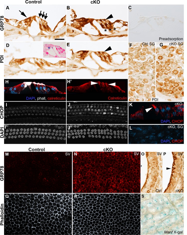
Fig. 4. Manf inactivation triggers ER stress and UPR activation in the cochlea.
Cochleas from 8-week-old Manfflox/flox;Pax2-Cre mice and control littermates were analyzed. Images were taken from the middle a–l or basal part of the cochlea m–s. a, b Transverse paraffin sections show ubiquitous GRP78 expression in the organ of Corti of control mice and GRP78 upregulation in cKOs. In OHCs, GRP78 is upregulated in the area below the cuticular plate (arrowhead). Large arrow marks IHC and small arrows OHCs. Deiters’ cells beneath OHCs are marked with stars. c GRP78 antibody was validated by preadsorption assay (see the section “Materials and methods”). d, e PDI shows also ubiquitous expression in controls and upregulation in cKOs. In OHCs, PDI upregulation has similar spatial characteristics (arrowhead) as GRP78. Inset in d is a reminder of Manf expression, shown by X-gal staining (see also Fig. 1). f, g Also SG neurons of cKO cochleas show PDI upregulation, revealed in paraffin sections. h, h′ The control whole mount is immunolabeled for calreticulin (red) and the maximum intensity projection is viewed in a transverse plane. Calreticulin expression pattern in OHCs (arrowheads) is comparable to GRP78 and PDI. Phalloidin (white) labels hair cell apices and pillar cells. DAPI labels nuclei (blue). i, i′, j, j′ Whole mount specimens show CHOP upregulation in OHCs of cKO cochleas. DAPI marks nuclei. k In the transverse section of a maximum intensity projection, CHOP expression (red) is seen in the first-row OHC. l Whole mount specimen from a cKO mouse shows absence of CHOP staining in SG neurons. DAPI marks nuclei (blue). m, n Stria vascularis whole mounts reveal GRP78 upregulation in marginal cells in cKO mice. o, p This upregulation in marginal cells (arrowhead) is also shown in paraffin sections. q, r Phalloidin labeling shows a regular hexagonal pattern of marginal cell boundaries, without signs of abrogated survival of these cells in cKO mice. s X-gal staining in a stria vascularis whole mount, viewed at the level of marginal cells, shows Manf expression (see also Fig. 1). Abbreviations: cKO conditional knock out; ctrl control; IHC inner hair cell; OHCs outer hair cells; ER endoplasmic reticulum; UPR unfolded protein response; SG spiral ganglion; GRP78 glucose-regulated protein 78; PDI protein disulfide isomerase; CHOP C/EBP homologous protein; SV stria vascularis. Scale bar (in a): a–e, h–j′, l 20 µm; f, g 25 µm; k 15 µm; m, n, q, r 37 µm; o, p 6 µm; s 12 µm.

