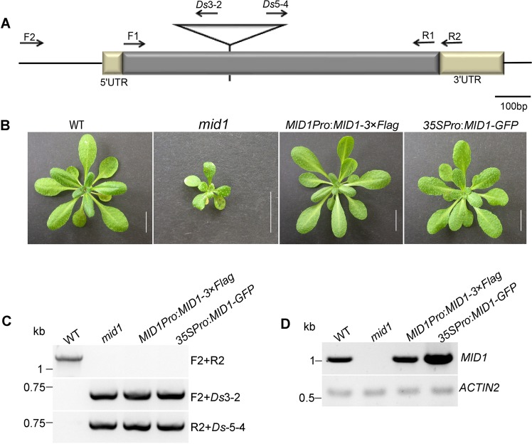
Figure 3.
Cloning of MID1. (A) Diagram of mid1 mutant. The Ds insertion is pointed out by a triangle. The position of primers used in (C,D) are displayed by arrows. (B) The phenotype of 4-week-old wild-type (WT), mid1, and genetically complemented lines MID1Pro:MID1-3 × Flag and 35SPro:MID1-GFP. Bar = 1 cm. (C) Genotyping of plants in (B) using the primers indicated in (A). (D) RT-PCR detection of MID1 cDNA in plants (B) to confirm the successful complementation. The primers F1 and R1 were used to RT-PCR analysis of MID1.

