
Figure 5.
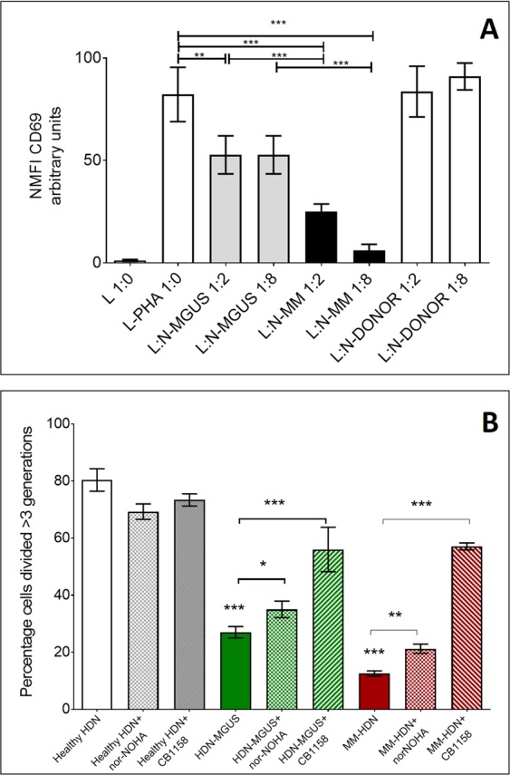
Arginase-1 confers both MGUS and MM high-density neutrophils immune suppressive properties. CD3 + T-cells obtained from healthy donors were activated using PHA and co-cultured with purified HDN freshly isolated from MGUS/MM patients or healthy donors, matched for sex and age, at increasing concentration (L:N ratio 1:2, 1:8). After 24 hours T-cells were examined for the early activation marker CD69. Bars represent the mean fluorescence intensity ± standard deviation of eight independent experiments, based on cells obtained from three different healthy donors, 6 MGUS and 6 MM/HDN (A). In an independent series of experiments, T-cells were labelled with CFSE and activated with PHA. After 3 hours, MGUS-HDN were added at the ratio L:HDNmgus 1:4; alternatively, MM-HDN were added at the ratio L:HDNmm 2:1 and cultured for 72 hours to measure proliferation. In the last 24 hours Arg-1 inhibitors nor-NOHA or CB1158 were added. Histograms show the percentage of proliferation of CFSE-labelled T-cells in presence of HDN and Arg-1 inhibitors (B). Stars denote p-value (***p < 0.001, **p < 0.05) using ANOVA test with post-hoc analysis. Abbreviations: L: lymphocyte, HDN: high-density neutrophils, PHA: phytohemagglutinin.