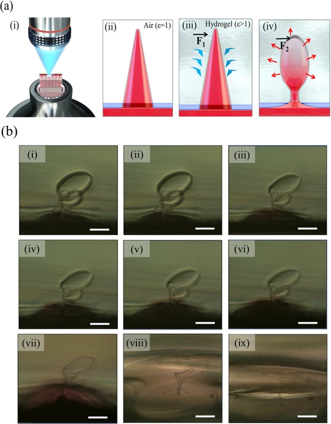
Figure 3.
(a) Schematic diagrams of (i) the dissolution of the HA microneedle in gelatin hydrogel and the needle dissolution mechanism; (ii) in air (ε = 1), (iii) absorption of moisture immediately after needle insertion in hydrogel, and (iv) dissolution of swollen needle (ε > 1). () indicates the absorption forces and () indicates the dissolution forces. (b) Optical images of HA microneedle dissolution over time in gelatin hydrogel; (i) immediately after needle insertion in hydrogel, (ii) after 2 min, (iii) 4 min, (iv) 6 min, (v) 8 min, (vi) 10 min, (vii) 30 min, (viii) 60 min, and (ix) 140 min. Scale bar = 200 μm.

