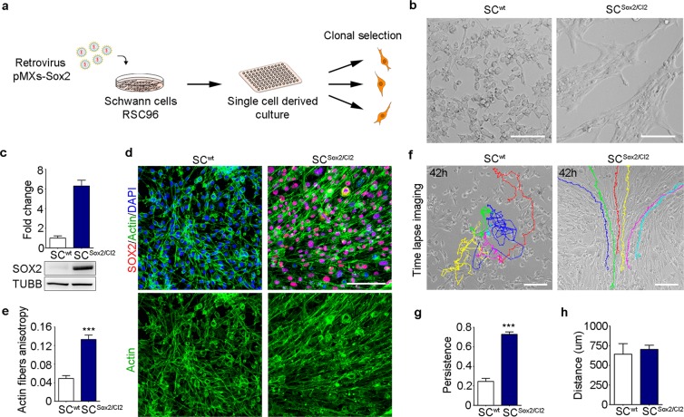
Figure 1.
Sox2 overexpression induces Schwann cell adhesion and directional migration. (a) Schematic representation of the protocol used to derive Sox2-positive clones from the rat Schwann cell line RSC96. (b) Bright-field images of SCwt and Sox2-positive clone SCSox2/Cl2 after Sox2 transduction. Scale bar, 100 μm. (c) Western blot analysis of total SOX2 protein levels in the whole lysate of SCwt and SCSox2/Cl2. TUBB was used as a loading control (N = 3). (d) Representative immunostaining confocal images of SOX2 (red) and Actin fibres (green) of SCwt SCSox2/Cl2 in FBS-supplemented medium. Nuclei were counterstained with DAPI (blue). Scale bar, 100 μm. (e) Quantification of actin fibre anisotropy by actin staining of SC and SCSox2/Cl2 (N = 5). (f) Representative time-lapse images from Supplementary Movies 1, 2 of SCwt and SCSox2/Cl2. Color lines show single-cell tracks of the Schwann cells for 42 hours. Scale bar, 100 μm. (g,h) Quantification of persistence and travelled distance of SCwt and SCSox2/Cl2, quantification was performed considering only 100 frames per cell (n = 10 cells). Graphs show mean value ± s.e.m, ***p < 0.0005.

