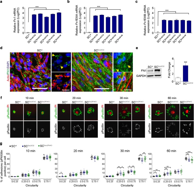
Figure 3.
Sox2 overexpression induces FN fibrillogenesis and focal adhesion in Schwann cells. (a–c) Graphs show real-time RT-qPCR measurements of Fn1, Fn1EIIIA and Fn1EIIIB mRNA levels in SCwt and selected clones after 3 days in culture. Gapdh, Ankrd27 and Rictor were used as reference genes, values are relative expression to SCwt. t-test comparing each clone with the control (N = 5). (d) Representative confocal images of Actin fibres (red) and FN1 (green) immunostainings showing FN expression and fibrillogenesis in SCwt (arrow head) an in SCSox2/Cl2 (arrows) cultured in FN-free medium. Nuclei were counterstained with DAPI (blue). Scale bars, 50 μm and 25 μm. (e) Western blot analysis and quantification of total FN1 protein in the whole lysate of SCwt and SCSox2/Cl2. GAPDH was used as a loading control. (N = 8). (f) pPaxillin immunostaining showing the formation of focal adhesion during the first 60 minutes after seeding on laminin-coated slides. (g) Distribution of adhesion structures during cell spreading, according to their circularity (n ≥ 25 cells/time point). Graphs show mean value ± s.e.m, *p < 0.05, **p < 0.005, ***p < 0.0005.

