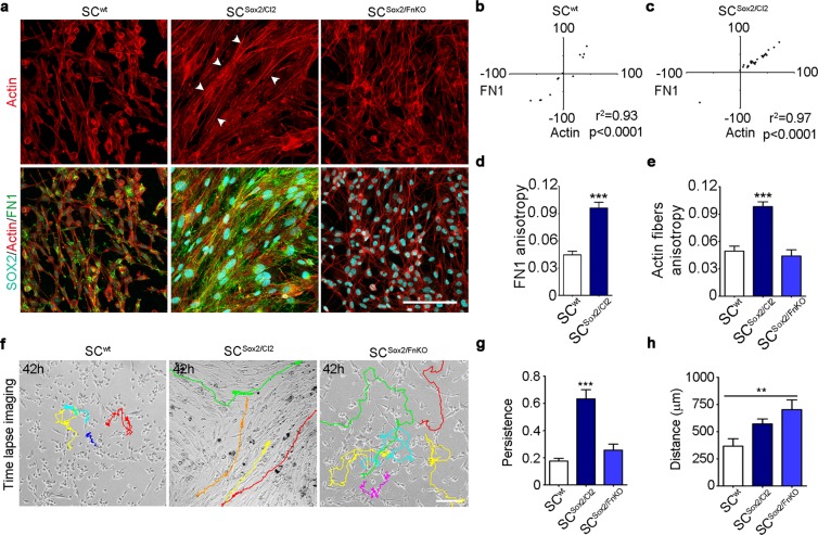
Figure 4.
Sox2-induced FN fibrillogenesis is responsible for Schwann cell organization and persistent migration. (a) Representative confocal images of SOX2 (cyan), Actin fibres (red) and FN1 (green) immunostainings of SCwt, SCSox2/Cl2 and SCSox2/FnKO. Arrow heads show the organization of the actin fibres in the SCSox2/Cl2. Scale bar 100 μm. (b,c) Correlation of FN1 fibre angles with the orientation of actin fibres in SCwt and SCSox2/Cl2 (N = 3) (d,e) Quantification of FN1 anisotropy of SCwt and SCSox2/Cl2 and actin fibres of SCwt, SCSox2/Cl2 and SCSox2/FnKO (N = 3, n ≥ 12 areas) (f) Representative time-lapse imaging from Supplementary Movies 5–7 from SCwt, SCSox2/Cl2 and SCSox2/FnKO respectively, cultured in KSRM (FN-free). Color lines show single-cell tracks of the Schwann cells during 42 hours. Scale bar, 100 μm. (g,h) Quantification of the distance and persistence of SCwt, SCSox2/Cl2 and SCSox2/FnKO migration in KSR medium, quantification was performed considering only 100 frames per cell (n = 10 cells). Graphs show the mean ± s.e.m. **p < 0.005, ***p < 0.0005.

