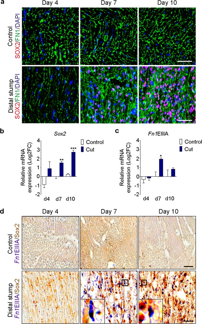
Figure 8.
Sox2 and FNEIIIA are up-regulated and co-expressed in Schwann cells after sciatic nerve transection. (a) Representative confocal images of FN1 (green) and SOX2 (red) immunostainings of rat sciatic nerves in control conditions or after complete transection, at day 4, 7 and 10 post surgery. Nuclei were counterstained with DAPI (blue). Scale bar, 50 μm. (b,c) Real-time RT-qPCR measurements of Sox2 and the Fn1 containing the spliced domain EIIIA from control and transected nerves at day 4, 7 and 10 post surgery. Ankrd27 and Rictor were used as reference genes, graphs show the mean value ± s.e.m. t-test comparing cut nerves with the controls in each time point, *p < 0.05 **p < 0.005, ***p < 0.0005. (N = 3) (d) In situ hybridization using a probe against Fn1EIIIA (purple) and SOX2 DAB staining (brown) in control and transected nerves at day 4, 7 and 10 post surgery. Insets in the bottom panels show the co-expression of Fn1EIIIA with a Sox2-positive Schwann cell at day 7 and 10 post surgery. Scale bar, 50 μm.

