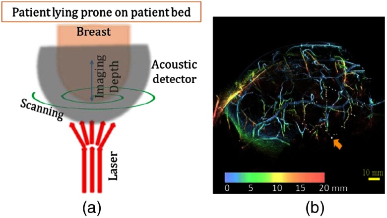
Fig. 3.
(a) Schematic of the hemispherical (HDA) breast imaging system. The breast is placed inside the HDA cup. The whole array is scanned in a spiral pattern along the horizontal plane. (b) PA MIP image of a healthy breast. The orange arrow corresponds to the deepest point from skin surface at 27 mm. This work by Toi et al.46 is licensed under a Creative Commons Attribution 4.0 International License.

