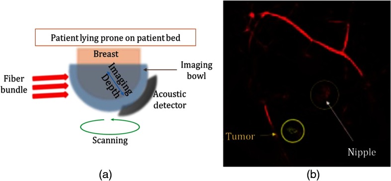
Fig. 4.
(a) LOUISA 3-D schematic and (b) MIP in sagittal view of a 3-D PA image showing small tumorous growth and microvessel-filled nipple. The 3.5-mm tumor is not conclusively ascertained. Therefore, this is used to demonstrate the sensitivity of LOUISA-3D and not its specificity if tumor differentiation. Reproduced with permission.51

