
Fig. 5.
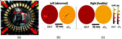
(a) The top view of fPAT breast interface. Red solid arrows indicate that the diameter of the breast interface is radially adjustable; red dashed arrows indicate the path of the laser beam. (b) An invasive mammary carcinoma with high-grade ductal carcinoma in situ (DCIS) in the left breast. Coronal HbT concentration and (oxygen saturation) maps of a breast with invasive mammary carcinoma with high-grade DCIS. The arrows indicate the suspicious lesion area. (c) Coronal HbT and maps for the contralateral healthy breast. © 2015 Am. Assoc. Phys. Med. Reproduced with permission.32