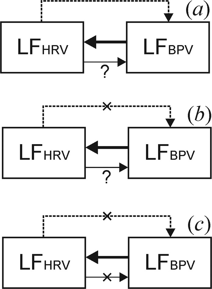
Figure 1.

Schematic representation of the structure of couplings between the LF components in the signals of the systems of HR and vascular tone regulation in the normal state (a) and during cardiac surgery in CPB without cardioplegia (b) and under cardioplegia (c). Arrows indicate the directions of couplings. The dashed line is a hemodynamic coupling from HRV to BPV, the solid line indicates an autonomous regulatory influence from BPV to HRV and the thin line indicates an autonomous regulatory influence from HRV to BPV.
