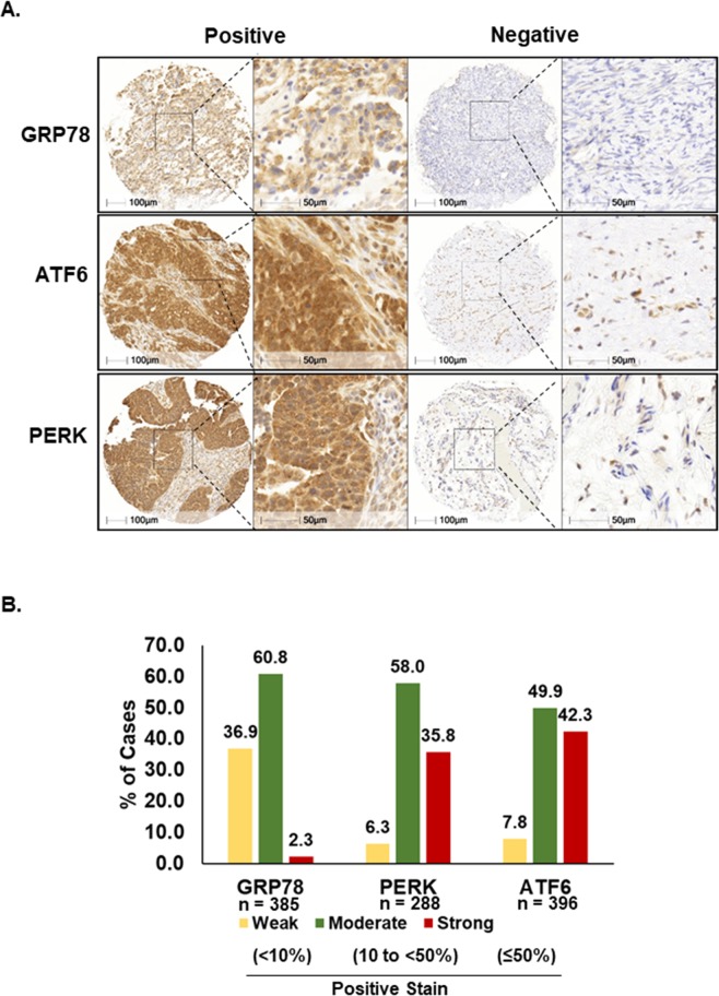
Figure 2.
Immunohistochemical expression patterns of ER stress-associated proteins in ovarian cancer tissues. (A) Representative examples of positive and negative/weak staining patterns of GRP78, ATF6, and PERK in ovarian cancer tissues. (B) Percent of cases of strong (≥50%), moderate (10 to <50%), and weak (<10%) expression for each protein are shown. Percent of staining was automatically quantified by HALO (Indica labs, New Mexico, USA).

