Figure 9.
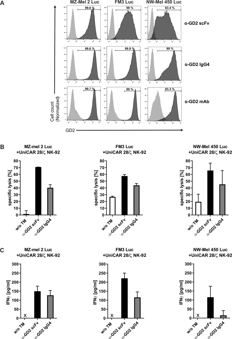
Specific cytotoxicity of UniCAR NK-92 cells redirected by α-GD2 TMs towards melanoma cells. (A) MZ-Mel 2 Luc, FM3 Luc and NW-Mel 450 Luc melanoma cells were incubated with α-GD2 scFv or α-GD2 IgG4 TMs. TM binding was then detected with antibody 5B9 specific for the E5B9 epitope tag, followed by Alexa Flour 647-conjugated goat α-mouse antibody (dark grey areas). As controls, cells were incubated with antibody 5B9 and secondary antibody without a TM (light grey areas). To confirm GD2 expression, the melanoma cells were also stained with an α-GD2 monoclonal antibody or an isotype-matched control antibody, followed by Alexa Flour 647-conjugated goat α-mouse antibody (bottom panels; dark grey and light grey areas, respectively). (B) UniCAR 28/ζ NK-92 cells were co-cultured at an E:T ratio of 5:1 with the different melanoma cell lines in the presence or absence of α-GD2 scFv or IgG4 TMs for 4 hrs. Thereafter, specific lysis was measured using a luminescence-based assay. Results are shown as mean ± SD of data from two independent experiments. (C) Supernatants collected from the cytotoxicity assays were analysed for the presence of IFNγ using an ELISA. Results are shown as mean ± SD for triplicate samples. x, not detectable.

