Figure 1.
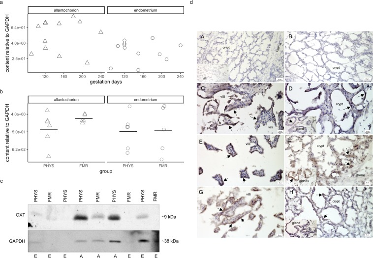
Oxytocin peptide content and immunolocalization. (a) Oxytocin peptide content relative to that of glyceraldehyde-3-phosphate dehydrogenase (GAPDH) during pregnancy. Protein content was quantified by western blotting with densitometry. Symbols correspond to tissues from individual mares (Δ, allantochorion; ○, endometrium). Note the logarithmic scales in panels a and b. (b) Oxytocin peptide content relative to that of GAPDH in physiological parturition (PHYS) and parturition with fetal membrane retention (FMR). Horizontal lines indicate geometric means. (c) Representative blots of oxytocin and GAPDH. Uncropped blots are presented in Supplementary Fig S4. Image Lab version 5.2.1 software was used for visualization and densitometry (Bio-Rad Laboratories, https://www.bio-rad.com/en-pl/product/image-lab-software?ID=KRE6P5E8Z). Abbreviations: OXT, oxytocin; A, allantochorion; E, endometrium. (d) Tissue expression and localization of oxytocin in horse placenta. Oxytocin peptide was visible as dark brown to black cytoplasmic staining (arrows). Images of negative controls (A,B): (A) endometrium with primary antibody omitted (no staining visible); inset in (A), allantochorion with primary antibody omitted (no staining visible); (B) endometrium stained with antibody incubated with blocking peptide (no staining visible). Images of the pregnancy group (C,D): (C) allantochorion with positively stained epithelial cells on villi; (D) endometrium with positively stained epithelial cells in crypts; inset in (D), endometrial glands with positively stained endothelial cells. Images of the physiological parturition group (E,F): (E) allantochorion with positively stained epithelial cells on villi; (F) endometrium with positively stained epithelial cells in crypts; inset in (F), endometrial glands with positively stained endothelial cells. Images of the fetal membrane retention group (G,H): (G) allantochorion with positively stained epithelial cells on villi; (H) endometrium with positively stained epithelial cells in crypts; inset in (H), endometrial glands with positively stained endothelial cells. Micrographs were made with Zen 2012 (blue edition) software (Zeiss, https://www.zeiss.com/microscopy/int/products/microscope-software/zen-lite.html).

