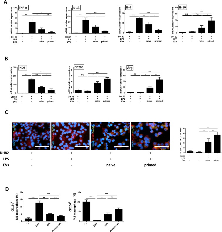
Figure 4.
EVs from primed cASCs induce the expression of M2 macrophage marker in vitro and in vivo. LPS-stimulated DH82 were co-cultured with EVs from naïve or primed cASCs for 48 h. (A) Relative mRNA expression levels of TNF-α, IL-1β, IL-6 and IL-10 in RAW 264.7 and DH82 cells. (B) Relative mRNA expression of iNOS, CD206 and Arg are shown. DH82+ : exist, LPS−: non-treated, LPS+ : treated, EVs−: absence. (C) Representative immunofluorescence staining using anti-CD11b-PE or anti-CD206-FITC positive cell, and the calculated percentage of CD206-FITC positive cells among the CD11b-PE positive cell are shown. (D) CD11c+M1 and CD206+M2 peritoneal macrophage in DSS induced colitis mice model. Data are shown as mean ± S.D. (ns = Not Statistically Significant *P < 0.05, **P < 0.01, ***P < 0.001 by one-way ANOVA analysis).

