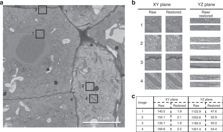
Fig. 3. An SBF-SEM section of an Arabidopsis thaliana root tip and four ROIs that show the denoising performance of DenoisEM.
a The original image and four annotated ROIs and b a qualitative comparison of the original and denoised cross sections. For each ROI we show an XY and YZ section to illustrate that the image quality also improves along the Z direction, even though DenoisEM restores each XY slice independently. c For each ROI, we provide an estimation of the noise standard deviation σn ref. 51 in the raw and denoised patch to illustrate the image quality quantitatively. Note that the noise level decreases by almost two orders of magnitude.

