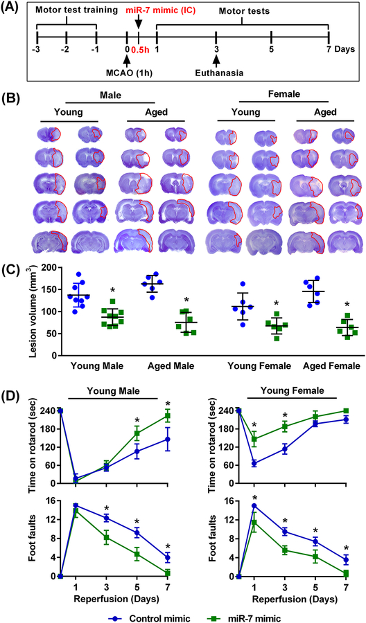
Fig. 3: Post-ischemic intracerebral treatment with miR-7 mimic protected the rat brain irrespective of age and sex.
(A) Schematic diagram of the experimental design, wherein rats were subjected to 60 min (1h) transient MCAO followed 30 min (0.5h) thereafter (during reperfusion) by injection of the miR-7 or control mimic. Brains were collected at 3 days of reperfusion for lesion volume assessment. Motor training was performed on separate cohorts of rats for 3 days prior to 60 min (1h) transient MCAO, followed 30 min (0.5h) thereafter (during reperfusion) by injection of the miR-7 or control mimic, and then motor testing at 1–7 days after. (B) Representative cresyl violet-stained serial sections (B) and lesion volume (C) in brains from the miR-7 mimic- and control mimic-treated groups of both sexes and ages. Lesion volume was measured at day 3 of reperfusion after 60 min transient MCAO. Data are mean ± SD (n = 6–9 rats per group). *p<0.05 compared to the control mimic group, by Mann-Whitney U test. (D) Recovery of functional performance assessed by the rotarod test and beam-walk test by young male and female rats that received an intracerebral injection of either control or miR-7 mimic 30 min after 60 min transient MCAO. The performance was assessed before treatment (time 0) and on days 1 through 7 of reperfusion. Data are mean ± SD (n = 4 rats per group). *p<0.05 compared to the respective control mimic group, by repeated measures ANOVA followed by Sidak’s multiple comparisons post-test.

