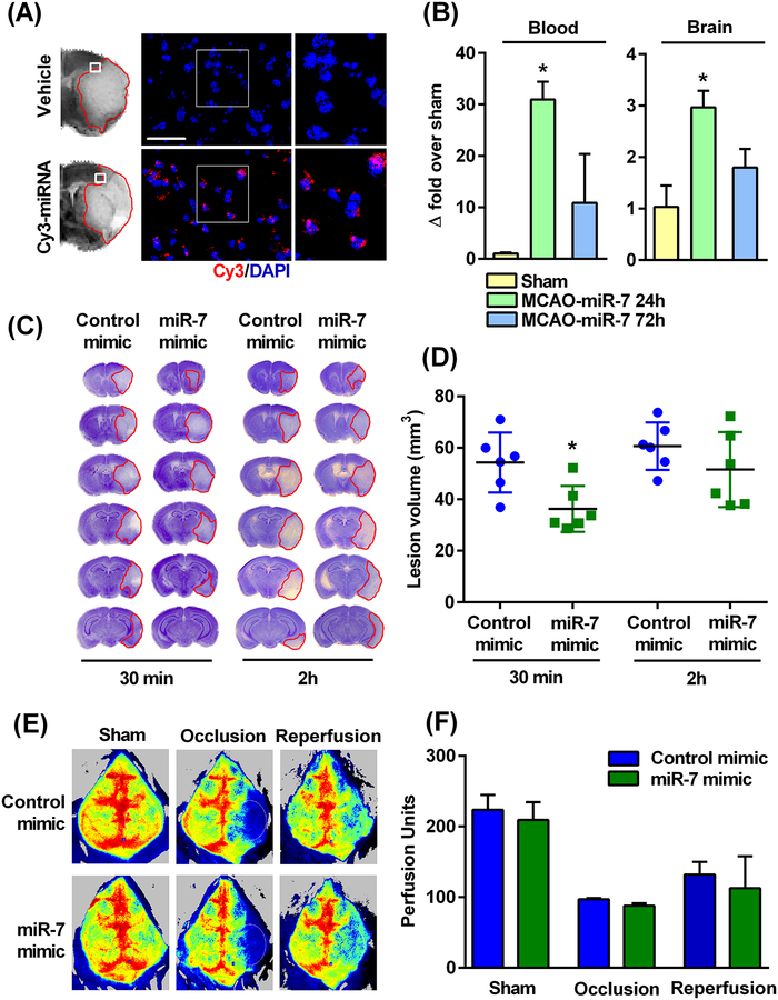
Fig. 4: Post-ischemic IV injection of miR-7 mimic decreased ischemic brain damage in young male mice.
(A) A Cy3-labeled mimic injected IV (retro-orbital venous sinus) at 30 min of reperfusion following 90 min transient MCAO was observed to be present in the peri-infarct region of the ipsilateral cortex at 24h of reperfusion. (B) The abundance of miR-7, assessed by real-time PCR, in blood and the peri-infarct region of the ipsilateral cortex of young male mice at 24h and 72h of reperfusion after 90 min transient MCAO, relative to those that underwent a sham (control) procedure. Data are mean ± SD (n = 4 mice per group). *p<0.05 compared to sham, by Kruskal-Wallis one-way ANOVA followed by Dunn’s post-test. (C and D) Representative cresyl violet-stained serial sections (C) and lesion volume (D) in brains from the miR-7 mimic- and control mimic-treated groups injected at either 30 min or 2h of reperfusion. Lesion volume was measured at day 3 of reperfusion after the 90 min transient MCAO. Data are mean ± SD (n = 6 mice per group). *p<0.05 compared to the control mimic group, by Mann-Whitney U test. (E and F) Representative in vivo laser speckle imaging (E) showing changes in cerebral blood flow before, during, and 24h after 90 min transient MCAO from the miR-7 mimic- and control mimic-treated groups injected at 30 min of reperfusion. Data are mean ± SD (n = 4 mice per group). *p<0.05 compared to the corresponding control mimic group, by Mann-Whitney U test (G). Scale bar, 30 μm.

