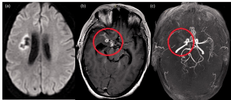
Figure 6.
Brain magnetic resonance imaging of a 35-year-old woman with a history of tubercular meningitis and vasculitis. (a) Diffusion-weighted image shows chronic infarct in the right centrum semiovale. (b) Axial T1 postcontrast image shows basal leptomeningeal enhancement and enhancing tuberculomas concentrated around the left middle cerebral artery (MCA) (circle). (c) Three-dimensional time-of-flight magnetic resonance angiogram shows typical narrowing involving the right supraclinoid internal carotid artery and total occlusion of the right MCA with nonvisualization of distal cortical branches (circle); normal-caliber posterior fossa vessels.

