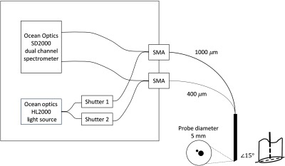
Fig. 2.

MDSFR system schematic: light from the source is split into two fibers and led to two shutters that guide the light either to the connector of the or the measurement fibers that first transport the light to the tissue and subsequently collect backscattered light from the tissue and guide it back to the spectrometer. The setup has two channels that are activated one-by-one. The two measurement fibers are integrated into a single-measurement probe. The operation of the setup is controlled by a LabView program (LabView, National Instruments, Austin, Texas).
