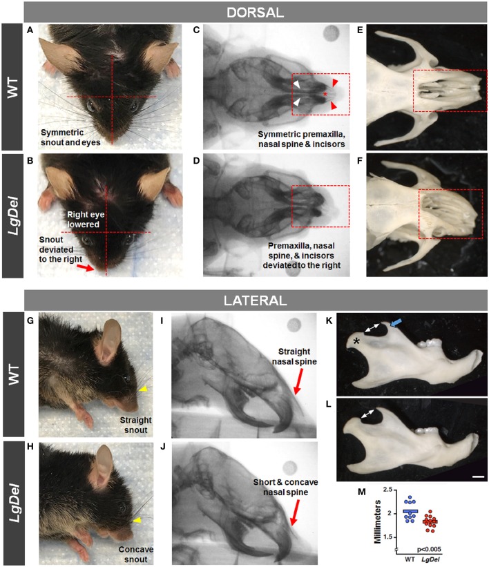
Figure 6.
Craniofacial anomalies in LgDel mice identified via facial photographs, skull radiographs, and bone morphology. Representative images of a WT mouse in dorsal and lateral view (A,G) and skull bones (C,E,I) showing symmetric facial features. Some LgDel mice had facial asymmetry involving the eyes and snout (B,H), and skull abnormalities involving the nasal spine, premaxilla, and incisors (D,F,J). The mouse depicted here displayed all of these abnormalities; however, this phenotype had low penetrance. Representative examples of right mandible from WT (K) and LgDel (L) mice showing difference in morphology of the coronoid process (blue arrow) and condyle (asterisk). (M) Quantification shows LgDel mice have a significantly shorter distance between the coronoid process and the head of the mandible than their WT counterparts (p < 0.005). Scale bar = 1 mm.

