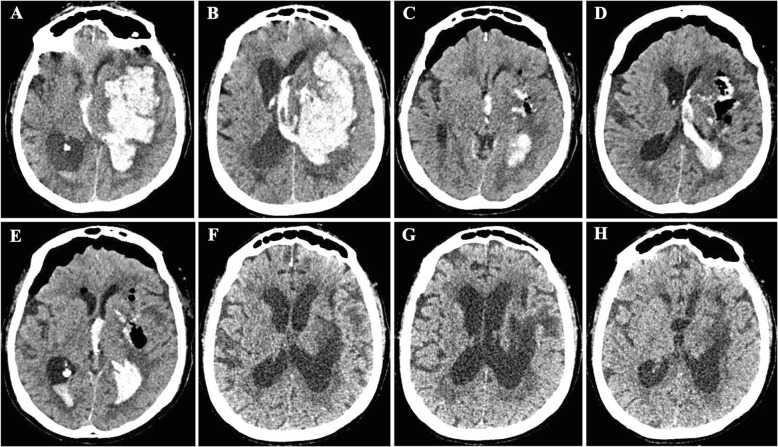
Fig. 3.
Case 02 of open craniotomy for hematoma drainage. a, b Day 1—Large hematoma in the left cerebral hemisphere leading to collapse of the left lateral ventricle with a midline shift of 12 mm, with a large left ventricular and third ventricle flooding, as well as diffuse effacement of cortical sulci of that hemisphere. c–e Day 2—Left frontoparietal craniotomy, with well-positioned bone fragment, aligned and fixed with metal clips. Reduction of the left frontal/frontotemporal intraparenchymal hematic content, with remnant hematic residues and air foci in this region. There was a significant reduction in the mass effect, with a decrease in lateral ventricular compression and a reduction in the midline shift. Bifrontal pneumocephalus causing shift and compressing the adjacent parenchyma. f–h Day 36—Resolution of residual hematic residues and pneumocephalus. Encephalomalacia in the left frontal/frontotemporal region. Despite the good surgical results, the patient remained in vegetative state

