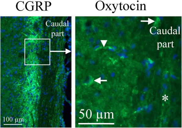
Fig. 10.

Oxytocin immunohistochemistry of the Spinal trigeminal nucleus. To the right, oxytocin immunohistochemistry is shown. A few cell bodies (arrows) express oxytocin in the Sp5. No immunoreactivity was found in fibers (asterisk). The arrowhead points at a stained capillary. On the left, CGRP expression in this region [25] is shown for comparison. The square in the left panel indicates the approximate location of the area presented in the right panel
