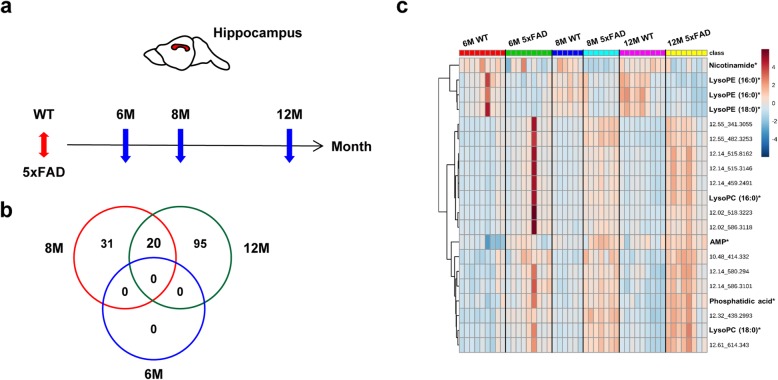
Fig. 1.
Hippocampal metabolomics of 5xFAD mice at different disease progression stages. a Time course of the hippocampal sample collection. b Venn diagram representing overlapping ion features that were significantly different between the hippocampi of WT and 5xFAD mice (q < 0.05) at 6, 8, or 12 months of age. c Hierarchically clustered heat map of the relative intensity of 20 metabolic markers. Rows and columns represent the individual mice and the 20 selected metabolites (retention time_m/z, *identified or putative metabolites), respectively. Each cell is colored based on the relative intensity

