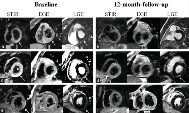
Figure 2.
Cardiac magnetic resonance findings in three different patients, at baseline (a, c and e) and at 12-month follow-up (b, d and f). Short-tau inversion-recovery images in a, c and e patients reveal hyperintense areas of myocardial edema (arrows). Early gadolinium enhancement images in a and c patients, showing early gadolinium enhancement areas (arrows, a, c, and e). At 12-month follow-up, a complete resolution of edema and hyperemia was recorded in all three patients (b, d and f) while areas of late gadolinium enhancement persisted (arrows). Cardiac magnetic resonance findings had a subepicardial to mid-wall distribution. STIR: Short-tau inversion-recovery, EGE: Early gadolinium enhancement, LGE: Late gadolinium enhancement

