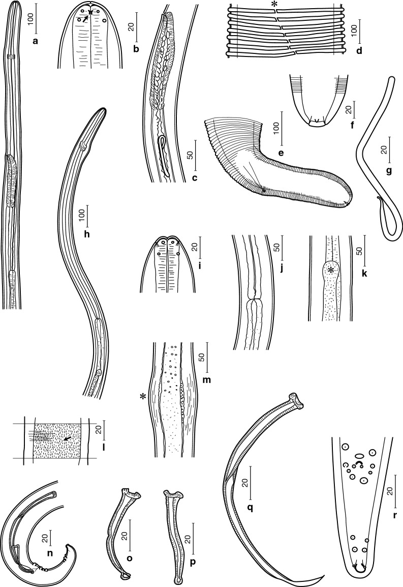
Fig. 1.
Line drawings of Onchocerca borneensis n. sp. Females (a–f), microfilaria (g) and males (h–r). a Anterior end, left lateral view. b Anterior extremity, lateral view, showing amphid (arrow). c Vagina, left lateral view. d Transverse cuticular ridges at midbody region, showing lateral field (*). e Posterior end, left lateral view. f Posterior extremity, ventral view, showing internal terminal point and two subterminal phasmids. g Microfilaria without sheath. h Anterior end, lateral view. i Anterior extremity, dorsoventral view. j Oesophago-intestinal junction. k Apex of testis (*). l Short longitudinal cuticular crests (arrow) at midbody region. m Body swelling (*). n Posterior end, right lateral view. o Right spicule, lateral view. p Right spicule, dorsoventral view. q Left spicule, lateral view. r Posterior end, ventral view. Scale-bars are in micrometres

