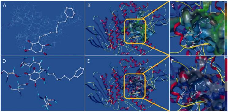
Figure 8.
Docking binding model of 15d with yeast α-glucosidase. (A): Binding mode of 15d docked with the prototype molecular of the active site. (B) and (C): Active site MOLCAD surface representation of lipophilic potential. (D): The interaction of 15d with the surrounding amino acids. (E) and (F): Active site MOLCAD surface representation of hydrogen bonding.

