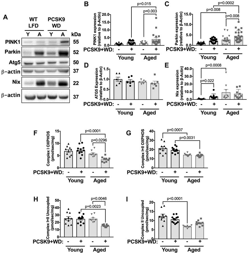
Figure 6. Aged but not young mice show reduced mitochondrial function and increased levels of Parkin and PINK1 during atherogenesis.
(A) Thoracic aortas were harvested from 3-month or 18-month WT, LFD-fed mice, and WT mice maintained on a low-fat diet until either 3-months or 18-months which were then transfected with PCSK9-AAV and fed a WD for 10 weeks. Lysates were blotted against PINK1, Parkin, ATG5, Nix and β-actin (Note, that Nix immunoblot was run separately to other proteins and has its own loading β-actin control, below). (B-E) Quantification of immunoblots from A. (F) Maximal thoracic aorta OCR with substrates for complex I OXPHOS. (G): Maximal aortic OCR with substrates for complex I+II OXPHOS. (H) Maximal aortic complex I+II uncoupled OCR. (I) Maximal aortic complex II uncoupled OCR. Each data point represents a biological replicate. Note, that the normolipidemic data (i.e., WT LFD denotation) are the same data shown in Figure 1. Data from Figure 1 and Figure 6 were run contemporaneously. All results are presented as mean ±SEM. 2-way ANOVA with Sidak’s post-hoc test for B-C and F-I. Kruskal-Wallis with Dunn’s post-hoc test for E. A=aged, Y=young.

