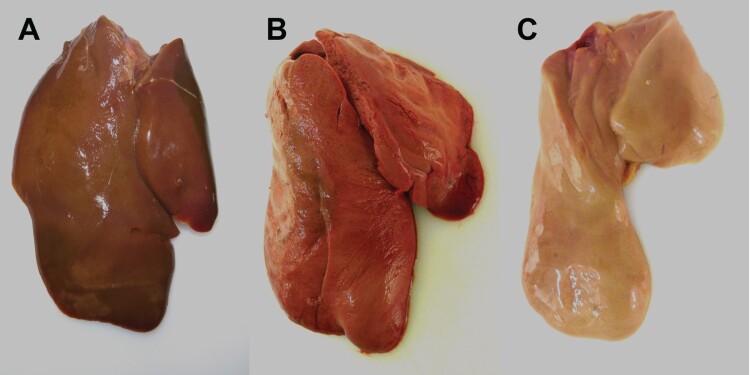
Figure 4.
Macroscopic findings in the livers of experimentally H5N8B-infected ducks. (A) Seronegative Pekin duck, H5N8B-infected, euthanized 9 dpi due to neurological symptoms, liver. Macroscopically normal, brown-red, acutely-angled liver without immunohistochemically-detectable hepatocellular influenza A virus matrix protein. (B) Pekin contact duckling in the mallard group, died 4 dpc, liver. Swollen, brick-red-colored, friable liver with rounded edges, interpreted as severe, acute, diffuse, necrotizing hepatitis with immunohistochemically-detectable hepatocellular influenza A virus matrix protein. (C) Seropositive mallard, H5N8B-infected, clinically normal, 14 dpi, liver. Swollen, beige, greasy liver with rounded edges, interpreted as moderate, acute, diffuse hepatocellular lipidosis (background pathology) without immunohistochemically detectable hepatocellular influenza A virus matrixprotein.

