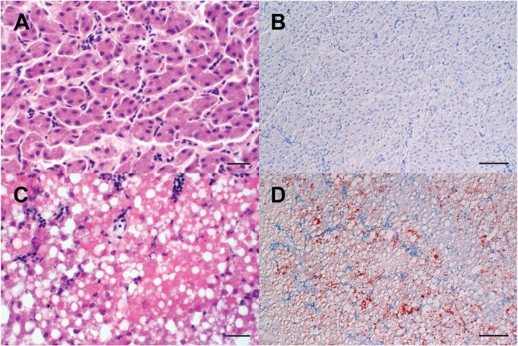
Figure 5.
Light microscopic finding in the livers of experimentally H5N8B-infected ducks. (A, B) Seropositive mallard, H5N8B-infected, clinically normal, 34 dpi, liver. (A) No obvious findings. (B) Lack of immunohistochemically-detectable hepatocellular influenza A virus matrixprotein antigen. (C, D) Pekin duck, contact animal, died 4 days post contact, liver. (C) Marked hypereosinophilia, hepatocellular vacuolation, membraneous rupture and nuclear pyknosis, karyorrhexis and lysis interpreted as severe, acute, coalescing to diffuse necrotizing hepatitis. (D) Immunohistochemistry reveals coalescing intrahepatocytic, intracytoplasmic and intranuclear influenza A virus matrix protein. (A, C) Hematoxylin-eosin, (B, D) Immunohistochemistry using the avidin-biotin-peroxidase-complex method with a monoclonal antibody against influenza A virus matrix protein (ATCC clone HB-64), 3-amino-9-ethylcarbazol chromogen (redbrown) and hematoxylin counterstain (blue). (A, C) bars = 20 μm. (B, D) bars = 50 μm.

