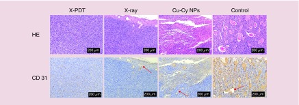
Figure 7. . X-ray-activated photodynamic therapy induced the inhibition of vascularization in squamous cell carcinoma tissue.
Histopathology examination stained by hematoxylin and eosin and immunohistochemistry examination stained by anti-CD31 antibody were used to assess the tumor vessels in squamous cell carcinoma tissue at day 16 after the treatments. CD31 positively stained tumor vessels in x-ray-activated photodynamic therapy (X-PDT), x-ray and copper-cysteamine nanoparticles groups were obviously less than control. Especially, there was nearly no tumor vessels observed in X-PDT group.
Cu-Cy NP: Copper-cysteamine nanoparticle; HE: Hematoxylin and eosin; X-PDT: X-ray-activated photodynamic therapy.

