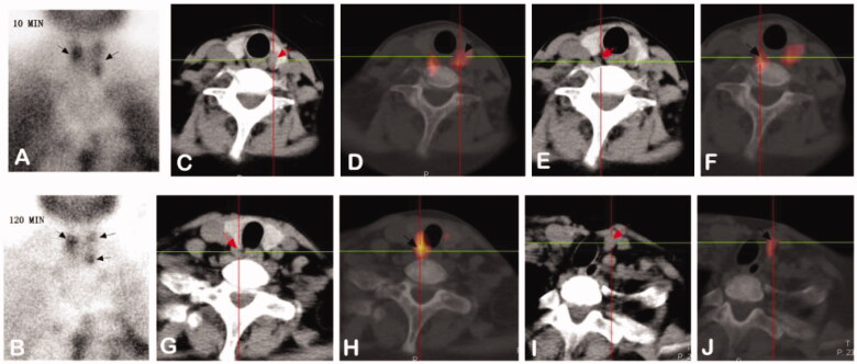
Figure 2.
Images from a 28-year-old female diagnosed with SHPT. (A,B): Dual phase 99mTc-MIBI planar scintigraphy of parathyroid glands. (A) early phase (after 10 minutes post injection of tracer) – showing radioactivity uptake in thyroid with increased distribution in left lobe and upper pole of right lobe. (B) late phase (after 120 min) – activity of wash-out from thyroid and retention in hyperfunctioning parathyroid glands (upper pole and inferior pole of left thyroid, upper pole of right thyroid, black arrows). (C–H): CT alone (C/E/G) and 99mTc-MIBI SPECT/CT images (D/F/H) of the same cross-section views showing the right upper (C and D), the left upper (E and F) and inferior (G and H) hyperfunctioning parathyroid glands (arrows). (I,J): CT alone (I) and 99mTc-MIBI SPECT/CT images (J) of the same cross-section views showing a new hyperfunctioning parathyroid gland that was not found in 99mTc-MIBI planar imaging in the inferior pole of right thyroid (arrows).

