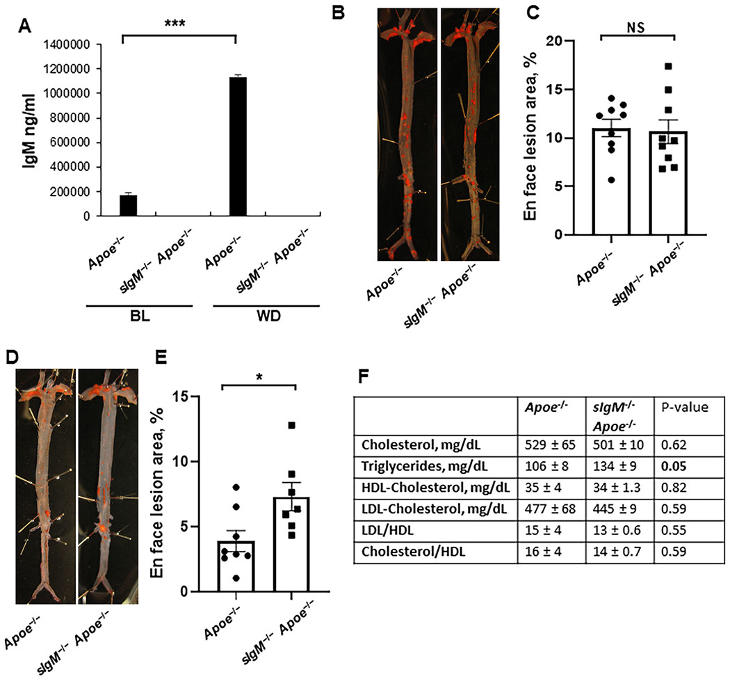
Figure 5. Low fat diet-fed sIgM deficient Apoe−/− mice exhibited enhanced atherosclerosis.
(A) IgM levels in serum of Apoe−/− and sIgM−/−Apoe−/− mice at baseline (BL) and after Western diet (WD) feeding. Values = mean ± SEM, n = 10 Apoe−/− BL, 10 sIgM−/− Apoe−/− BL, 7 Apoe−/− BL, 7 sIgM−/−Apoe−/−. (B, C) Sudan IV en face staining (B) and quantification (C) of lesion area in whole aorta of Apoe−/− and sIgM−/− Apoe−/− mice after 12 weeks of WD feeding. Values = mean ± SEM. (D,E) Sudan IV en face staining (D) and quantification (E) of lesion area in the whole aorta of standard laboratory diet-fed, 40 weeks old Apoe−/− and sIgM−/−Apoe−/− mice. Values = mean ± SEM. *P < 0.05 and ***P < 0.001, data analyzed by Student’s t-test. (F) Serum lipid profile analysis in standard laboratory diet-fed, 40 weeks old Apoe−/− and sIgM−/− Apoe−/− mice.

