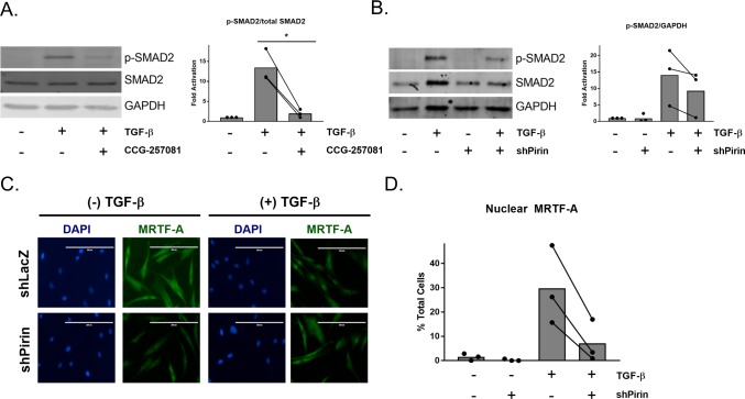
Figure 5.
CCG-257081 and modulation of pirin decrease SMAD2 phosphorylation and MRTF-A nuclear localization. (A) Left panel: Representative Western blot of p-SMAD2, total SMAD2, and GAPDH of lysates from primary human primary dermal fibroblasts after cotreatment with 10 μM CCG-257081 and 10 ng/mL TGF-β for 24 h. Right panel: Quantification of left panel. The results are expressed as the overall mean as well as mean values from three independent experiments. *, p < 0.05 using a ratio paired t test. (B) Human primary dermal fibroblasts were infected with virus containing shRNA against LacZ (shPirin −) or Pirin (shPirin +). Cells were then treated with 10 ng/mL TGF-β for 24 h, and Western blotting was performed for p-SMAD2, total SMAD2, and GAPDH. Right panel: quantification of left panel. Bars represent overall mean with paired independent means, n = 3. (C) Cells infected with shLacZ or shPirin were plated in 8-well chamber slides and then were treated with 10 ng/mL TGF-β in 0.5% FBS+DMEM and stained for endogenous MRTF-A. Scale bar represents 200 μm. (D) Quantification of (C) with the percentage of total cells with exclusively nuclear MRTF-A, n = 3. The overall mean is shown (bars). Total number of cells counted in each experimental condition was >200.

