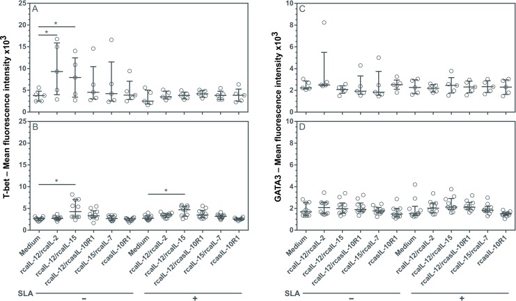
Fig 3. Evaluation of T-bet and GATA3 expression in lymphocytes from healthy and diseased dogs after stimulation with recombinant canine proteins.
PBMCs from healthy negative control dogs (n = 5) (A, C) and dogs with leishmaniasis (n = 10) (B, D) were cultured in medium alone (Medium) or medium containing rcaIL-12/rcaIL-2, rcaIL-12/rcaIL-15, rcaIL-12/rcasIL-10R1, rcaIL-15/rcaIL-7, or rcasIL-10R1 alone, with or without SLA. After 5 days, PBMCs were labeled anti-human T-bet FITC-conjugated antibodies, and anti-human GATA3 PE-conjugated antibodies or FITC-conjugated and PE-conjugated isotype control antibodies. Lymphocyte mean fluorescence intensity (MFI) was assessed by flow cytometry. Bars represent MFI median values and 25th and 75th percentile interquartile range. Symbols represent data of individual animals. Asterisks indicate significant differences (Friedman’s test with Dunn’s multiple comparison, p < 0.05).

