Figure 2.

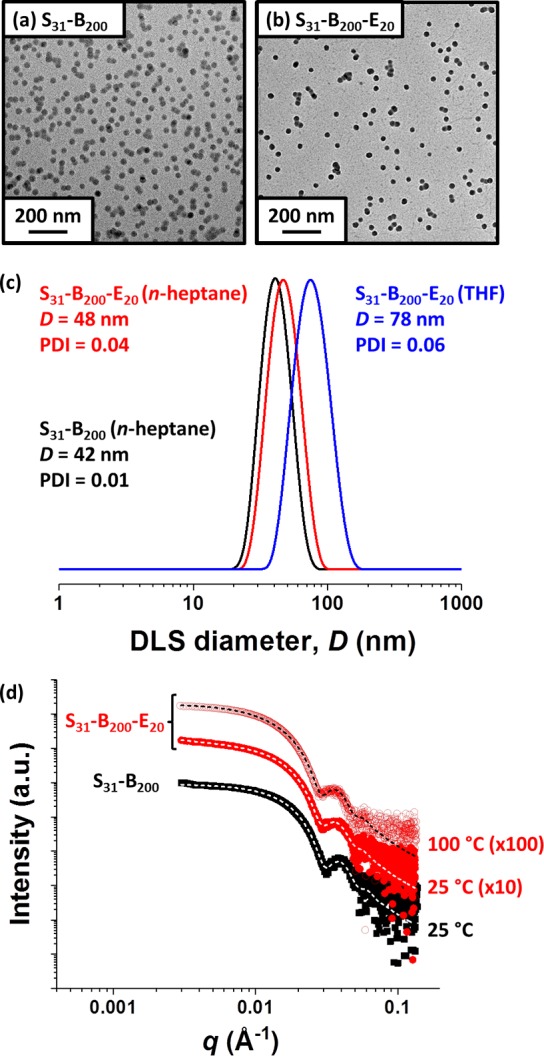
Transmission electron micrographs recorded for (a) linear poly(stearyl methacrylate)–poly(benzyl methacrylate) (S31–B200) spheres and (b) core cross-linked poly(stearyl methacrylate)–poly(benzyl methacrylate)–poly(ethylene glycol dimethacrylate) (S31–B200–E20) spheres. (c) DLS particle size distributions obtained for 0.10% w/w dispersions of linear S31–B200 spheres prepared using n-heptane as diluent (black data) and core cross-linked S31–B200-E20 spheres prepared using either n-heptane (red data) or THF (blue data) as diluent. (d) SAXS patterns recorded for 1.0% w/w dispersions of linear S31–B200 spheres in mineral oil at 25 °C (black squares) and core cross-linked S31–B200-E20 spheres in mineral oil at 25 °C (red circles) and 100 °C (open red circles). Dashed lines represent data fits using an established spherical micelle model.35 For clarity, SAXS patterns are offset by an arbitrary factor, as indicated.
