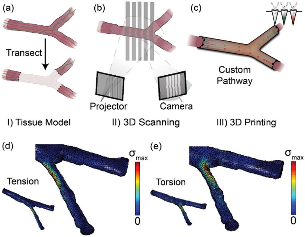
Figure 3.

(a)-(c) Personalized nerve guidance pathways via a combination of 3D scanning and 3D printing. The three critical steps are: (a) transection of the nerve tissue model, (b) imaging transected tissue, and (c) 3D printing scaffolds containing path-specific biochemical gradients. (d-e) Computational analysis of the nerve pathways via FEA simulation. Visualizing von Mises stress (σ) distribution in the nerve pathway under both (d) tensile and (e) torsional loading conditions applied to the distal ends of the nerve can be useful in providing an insight to the outcomes from in vivo studies and identifying areas requiring reinforcement. Reproduced with permission.[17] Copyright 2015, Wiley-VCH.
